
Unlocking the Secrets of Brain Aging—One Organoid at a Time
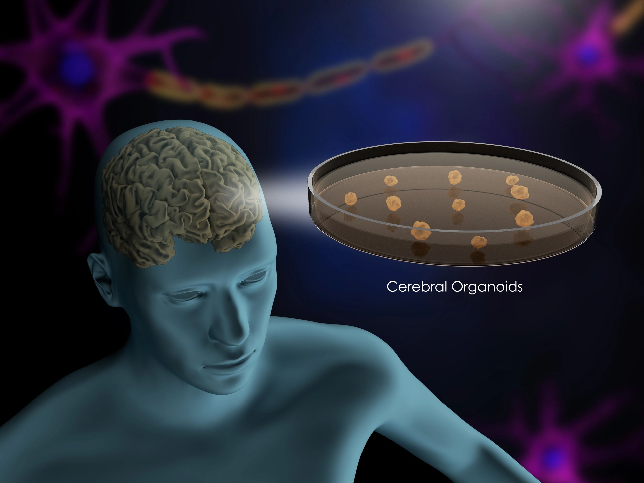
Imagine growing a miniature version of the human brain in a lab—complete with functioning neurons, complex circuitry, and the ability to model diseases—all without risking a single patient’s health. That’s the promise of brain organoids, 3D clusters of lab-grown neural tissue derived from stem cells that mimic key structural and functional features of the developing brain. These cutting-edge models are revolutionizing neuroscience by offering unprecedented access to brain development, neurological disorders, and now, the aging process.
Studying aging in traditional models has long been limited by species differences, ethical constraints, and the slow progression of age-related changes. Brain organoids overcome many of these hurdles, enabling researchers to observe cellular aging in real time and under controlled conditions. For families in Jhelum, Lahore, and beyond who face rising rates of neurodegenerative conditions like Alzheimer’s, this research could one day lead to earlier diagnostics and targeted therapies.
In this post, we’ll explore how scientists are using long-term organoid cultures to track age-related brain changes, what early findings reveal about cognitive decline, and how these models are accelerating the development of interventions that could preserve brain health across the lifespan.
Peering into the Future: What Brain Organoid Aging Means for Neurological Research
Recent breakthroughs in neuroscience are reshaping how researchers understand brain development and degeneration—with brain organoids now offering a dynamic model for studying cellular aging in real time. These 3D cultures, grown from stem cells, mimic early human brain structures and allow scientists to observe neural changes over extended periods. Unlike static tissue samples, organoids enable longitudinal tracking of synaptic activity, gene expression shifts, and protein aggregation—key markers linked to age-related neurological conditions such as Alzheimer’s and Parkinson’s.
For research teams across academic and clinical settings in Jhelum, Lahore, and beyond, these models provide a scalable platform to test interventions aimed at slowing cognitive decline. Observing aging patterns in organoids helps identify biological clocks within neurons, assess oxidative stress responses, and evaluate how environmental factors accelerate or delay degeneration. This data is crucial for developing targeted therapies that address root causes rather than symptoms.
Practical applications include:
- Validating drug efficacy on human-derived neural tissue before animal or clinical trials
- Mapping epigenetic changes associated with prolonged neural activity
- Simulating disease onset in controlled environments to improve diagnostic timelines
By integrating organoid studies with regional biobanks and collaboration networks, laboratories can enhance reproducibility and expand access to cutting-edge neuroscience tools—accelerating discoveries that matter most to aging populations.
5 Key Signs of Neural Aging Observed in Long-Term Brain Organoid Studies
Recent advancements in neuroscientific research have uncovered critical insights into how brain tissue ages over time—using lab-grown brain organoids as a model system. These 3D mini-brain structures, derived from stem cells, offer a window into the progression of age-related neurological changes without relying on animal or human subjects. Over extended culture periods, researchers have identified several consistent markers of cellular aging and neural decline. Below are the key findings that are reshaping how scientists understand neurodegeneration and cognitive deterioration.
-
Accumulation of Senescent Cells
As organoids mature past six months in culture, a growing number of cells enter a senescent state—they stop dividing but fail to die off. These lingering cells secrete inflammatory factors that disrupt surrounding neural networks, mirroring processes seen in aging brains and linked to conditions like Alzheimer’s. -
Decline in Synaptic Density
Over time, there’s a measurable reduction in synaptic proteins such as PSD-95 and synaptophysin, indicating fewer functional connections between neurons. This synaptic thinning parallels cognitive decline observed in older adults and may serve as an early biomarker for neurodegenerative risk. -
Increased Oxidative Stress and Mitochondrial Dysfunction
Aged organoids show elevated levels of reactive oxygen species (ROS) and reduced mitochondrial efficiency. This imbalance impairs energy production in neurons, contributing to slower signaling and increased vulnerability to damage. -
Emergence of Tau Protein Aggregates
Hyperphosphorylated tau—a protein associated with neurofibrillary tangles in Alzheimer’s—begins to accumulate in longer-cultured organoids. These aggregates disrupt axonal transport and precede visible neuron loss. -
Reduced Neuroplasticity Markers
Expression of genes tied to neuroplasticity, such as BDNF and CREB, diminishes significantly in older organoids. This suggests a declining capacity for learning and adaptation, similar to changes seen in aging human brains.
These findings help researchers screen potential therapeutic interventions aimed at slowing neural aging—all within a controlled, scalable model system.
Unlocking New Frontiers in Brain Health Research
The recent breakthrough in long-term brain organoid aging offers a transformative lens for understanding neurodegenerative conditions like Alzheimer’s and Parkinson’s. By replicating the aging process in lab-grown neural tissue, researchers can now observe age-related cellular changes in real time, providing unprecedented access to the biological mechanisms driving cognitive decline. This advancement allows scientists to test preventive therapies and monitor disease progression without relying solely on animal models or post-mortem human tissue.
For families in Punjab and beyond who face increasing risks of dementia, this research brings hope for earlier diagnosis and more targeted treatments. Researchers are now able to:
- Track protein accumulation and neuronal decay across simulated years of aging
- Identify genetic and environmental triggers that accelerate brain aging
- Validate drug candidates for neuroprotection in human-like neural environments
These models also support personalized medicine by integrating patient-derived cells, making it possible to predict individual responses to treatment. While still in early stages, such innovations lay the groundwork for preventative brain health strategies. Proactively managing cognitive wellness through lifestyle adjustments and early screening—especially for those with family histories of neurodegenerative diseases—can complement these scientific advances. As urban centers like Lahore and Jhelum expand access to neurological care, integrating new research insights into community health planning will be key to building resilient aging populations.
Your Questions Answered: What Brain Organoids Reveal About Human Aging
What exactly are brain organoids and how are they developed?
Brain organoids are 3D, miniaturized models of brain tissue grown from human stem cells in a lab environment. Scientists guide these stem cells—often derived from skin or blood samples—to differentiate into various neural cell types, self-organizing into structures that mimic early brain development. Using specialized culture conditions and bioreactors, researchers can sustain these organoids for months or even years, allowing them to study complex neurological processes in a controlled setting.
How do brain organoids mimic the human brain?
While not full-scale brains, organoids replicate key architectural and functional features of developing brain regions, such as cortical layering, neural network activity, and synaptic signaling. They express many of the same genes and proteins found in human brain tissue, making them valuable for modeling neurodevelopmental and neurodegenerative processes—especially as they mature over time.
Can brain organoids truly model aging?
Recent advances show that long-term cultured organoids develop aging-related markers, including oxidative stress, mitochondrial dysfunction, and accumulation of misfolded proteins—hallmarks also seen in aging human brains. These changes emerge spontaneously over extended culture periods, offering a window into how aging mechanisms unfold at the cellular level.
What are the limitations of using organoids to study aging?
Organoids lack blood vessels, immune cells, and sensory input, limiting their ability to replicate systemic aging. They also don’t represent late-life brain states perfectly, as they follow an intrinsic developmental clock. However, researchers are refining protocols to extend culture longevity and introduce stressors that accelerate age-like changes.
How can this research impact future treatments?
By identifying early molecular shifts tied to brain aging, scientists can test drugs or gene therapies aimed at delaying cognitive decline. These models are especially useful for studying conditions like Alzheimer’s and Parkinson’s in genetically diverse populations.
Are ethical concerns involved in brain organoid research?
Yes—while current organoids are far from conscious, their increasing complexity raises questions about neural sentience. Rigorous oversight ensures responsible innovation, balancing discovery with ethical boundaries.
What This Brain Organoid Research Means for the Future of Neurological Science
The emerging ability to observe long-term aging processes in brain organoids marks a pivotal shift in how scientists study neurodegenerative conditions like Alzheimer’s and Parkinson’s. These lab-grown, three-dimensional neural models offer an unprecedented window into human brain development and deterioration outside the body, enabling researchers to track cellular changes over time with remarkable precision. Unlike animal models, brain organoids derived from human stem cells reflect more accurate genetic and physiological responses, making them invaluable for early-stage drug screening and disease modeling. This breakthrough paves the way for identifying biomarkers of aging and testing therapeutic interventions years before symptoms appear in real patients. For clinicians and biotech innovators, this means faster translation from theory to treatment—and more targeted approaches to preserving cognitive health.
To leverage these advancements, consider:
- Supporting research initiatives that integrate organoid models into preclinical pipelines
- Exploring collaborations with neuroscience labs utilizing long-term culture techniques
- Staying informed about regulatory frameworks shaping ethical use of neural models
This isn’t just lab curiosity—it’s the foundation of next-generation neurological care. By embracing these tools, the medical and biotech communities can accelerate the development of anti-aging therapies and bring personalized neuroprotection closer to reality. The future of brain health starts in the dish.
Discover more from UnzipWorld: DIY, E-commerce, Fashion & Tech Insights
Subscribe to get the latest posts sent to your email.